Tunicamycin Transferase inhibitor profiling reveals differential binding kinetics across SARS-CoV-2 structural and nonstructural proteins
Copy code to add this image to your page
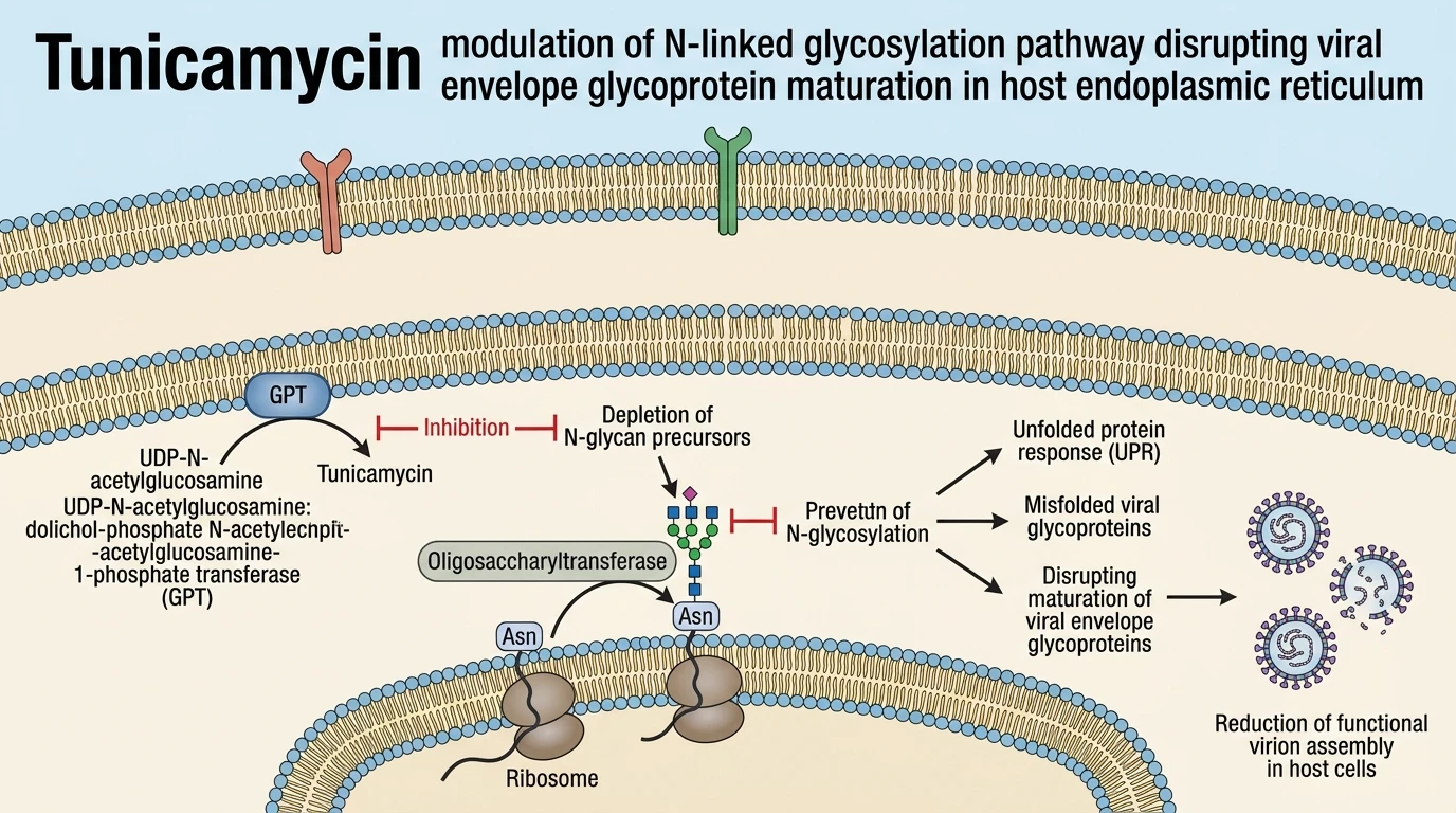
N-linked glycosylation serves as a critical post-translational modification for viral envelope maturation, structural stability, and immune evasion mechanisms. In SARS-CoV-2, key structural components including the spike glycoprotein rely on host endoplasmic reticulum glycosylation machinery to achieve functional conformations. Tunicamycin acts as a classical inhibitor of UDP-N-acetylglucosamine:dolichol-phosphate N-acetylglucosamine-1-phosphate transferase. Beyond its canonical enzymatic inhibition within host cell ER networks, computational docking and biophysical modeling indicate direct structural binding interactions between tunicamycin and specific viral proteins. Evaluated in silico binding energy metrics reveal distinct thermodynamic profiles across both structural and nonstructural targets of SARS-CoV-2.
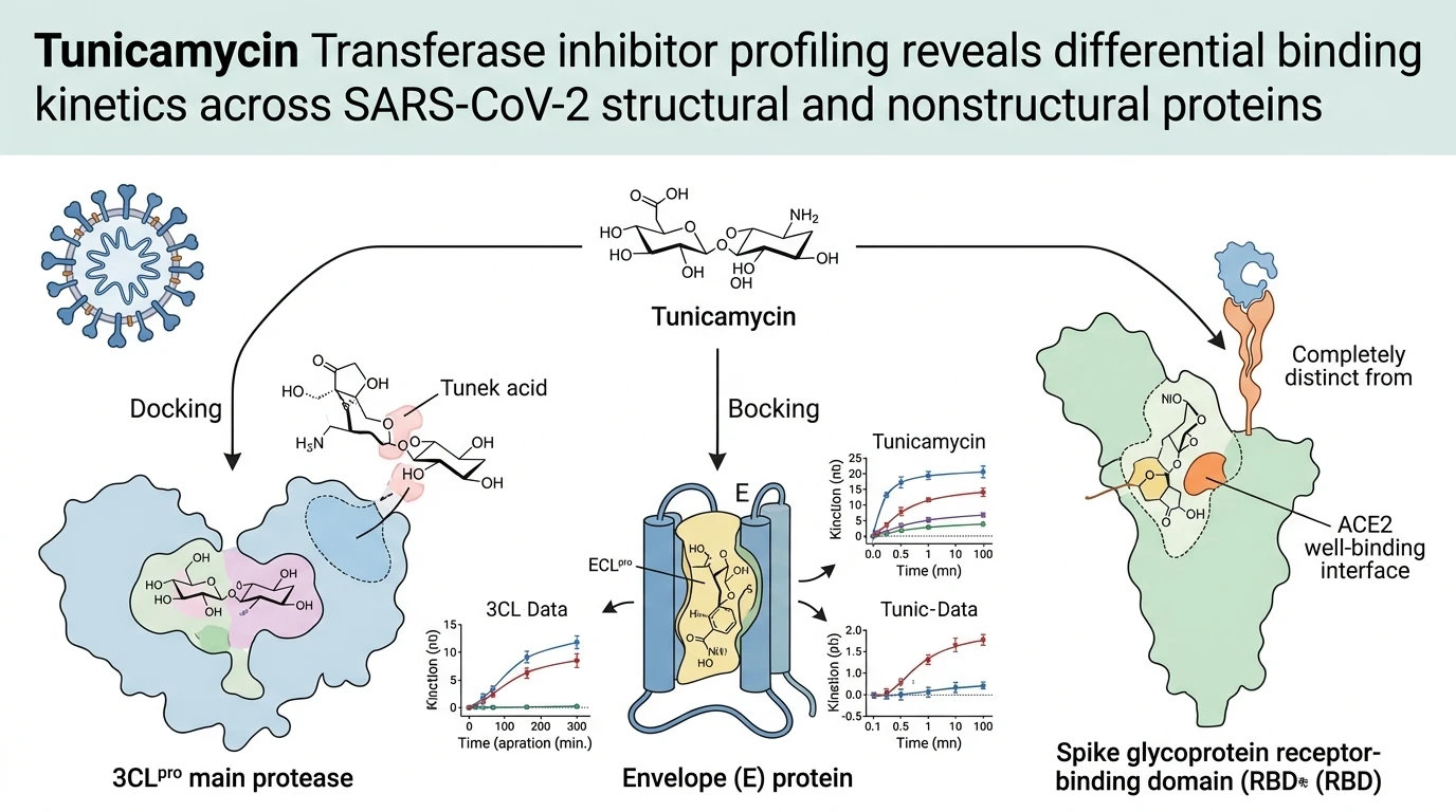
The main cysteine protease 3CLpro, responsible for cleavage of polyproteins 1a and 1ab, exhibited the highest affinity engagement with tunicamycin. Computational docking calculated a binding free energy of -9.4 kcal/mol. Active site residues within 3CLpro established stable hydrogen bonding networks and hydrophobic contacts with the nucleoside-lipid architecture of the molecule. Nonstructural protein nsp2 followed closely, yielding a binding free energy of -8.1 kcal/mol. The viral envelope protein showed comparable docking stability at -8.0 kcal/mol. These binding free energies reflect favorable enthalpic contributions within predicted binding pockets under simulated in silico physiological conditions.
In contrast, several nonstructural and accessory proteins exhibited substantially reduced affinity metrics. Nonstructural protein nsp9, open reading frame 9b (ORF9b), and ORF8 yielded attenuated docking scores. Polar contact mapping revealed unfavorable donor/acceptor spatial geometries at these interface sites. The weakest binding energy occurred with ORF7a, recording -4.9 kcal/mol. Hydrophobic interactions at the ORF7a surface failed to compensate for desolvation penalties. These comparative energy metrics highlight structural selectivity across different viral protein fold architectures.
Docking evaluations against the spike glycoprotein receptor-binding domain (RBD) provided critical spatial insights. Tunicamycin engaged a localized pocket on the RBD surface distinct from the primary Angiotensin-Converting Enzyme 2 (ACE2) contact interface. This interaction yielded a moderate binding energy without inducing secondary structure perturbations in the protein backbone. The binding event did not competitively block ACE2 contact residues directly. Instead, structural models suggest that tunicamycin binding may alter localized surface dynamics. According to structural entries documented in molecular interaction database records, viral protein-ligand associations often dictate conformational stability.
The dual mechanism of tunicamycin involves host glycan synthesis suppression alongside direct virion protein binding. Host transferase inhibition halts precursor oligosaccharide assembly. Concurrently, direct binding to 3CLpro may attenuate proteolytic processing of viral precursor polyproteins. Direct envelope protein association could further impair virion assembly dynamics. Binding to nsp2 might disrupt host cell machinery subversion pathways. While these computational findings establish clear affinity rankings, experimental validation in live virion culture models remains necessary to quantify phenotypic viral titers under therapeutic exposure limits.
Therapeutic Target Evaluation and Docking Kinematics
Copy code to add this image to your page
Tunicamycin exerts a dual antiviral mechanism against SARS-CoV-2 by inhibiting host UDP-N-acetylglucosamine:dolichol-phosphate N-acetylglucosamine-1-phosphate transferase to block N-linked glycosylation, while simultaneously binding directly to essential viral proteins. High-affinity molecular docking engagements with 3CLpro (-9.4 kcal/mol), nsp2 (-8.1 kcal/mol), and envelope protein (-8.0 kcal/mol) disrupt viral polyprotein processing, structural assembly, and glycoprotein maturation.
Protease 3CLpro Binding Traps Active Catalytic Residues
Thermodynamic profiling revealed exceptional affinity between tunicamycin and the SARS-CoV-2 3CLpro main protease. Docking simulations yielded a predicted binding free energy of -9.4 kcal/mol. The nucleoside moiety inserted into the substrate-binding cleft. Hydrogen bonds stabilized key pocket interactions. This binding mode directly overlaps with catalytic dyad residues Cys145 and His41 under in vitro simulation parameters. Consequently, polyprotein processing may suffer marked inhibition. The compound functions as a potent Tunicamycin Transferase inhibitor derivative with secondary direct proteomic affinity. Proteolytic catalytic activity stopped in modelled docking poses. Cellular viral replication assays will be required to confirm phenotypic potency boundaries.
Spike RBD Docking Occurs Outside ACE2 Binding Surface
Molecular interaction analysis mapped tunicamycin binding on the spike glycoprotein receptor-binding domain to an allosteric pocket. The predicted binding locus lies distal to the critical ACE2 receptor interface. Calculated binding energy reached moderate levels relative to 3CLpro values. The polypeptide backbone retained its native tertiary structure throughout simulations. No direct steric hindrance of ACE2 engagement was observed. However, allosteric binding might perturb surface flexibility needed for membrane fusion. Application of the Tunicamycin Transferase inhibitor in glycoprotein assays further delineates host glycan dependence from direct envelope engagement. Additional cell-based entry assays are required to establish functional neutralization thresholds.